Shape as Destiny: How Steroid Molecular Architecture Dictates Receptor Binding and Physiological Outcome
Photo: Gpruett2, CC BY-SA 3.0, via Wikimedia Commons
In pharmacology, few relationships are as elegant—or as consequential—as the one between a steroid molecule and its receptor. At the most fundamental level, every hormonal signal that a steroid transmits depends not on its chemical formula alone, but on the precise three-dimensional contours it presents when it arrives at a binding site. The difference between a compound that reduces life-threatening inflammation and one that reshapes muscle tissue can sometimes be traced to the repositioning of a single hydroxyl group or the addition of a methyl substituent at a specific carbon position. Understanding why requires a close look at the molecular mechanics of steroid-receptor interaction.
The Lock-and-Key Model Revisited
Early pharmacologists described drug-receptor interactions using the metaphor of a lock and key—a structurally specific ligand fitting into a complementary binding pocket. While that model remains instructive, modern structural biology has revealed a more dynamic picture. Nuclear receptors, the class of intracellular proteins that recognize most steroid hormones, do not simply accept or reject a ligand based on geometric fit. They undergo conformational changes in response to binding, essentially reshaping themselves around the incoming molecule.
This induced-fit mechanism has major consequences for how scientists interpret structure-activity relationships. When a glucocorticoid such as cortisol occupies the glucocorticoid receptor's ligand-binding domain, it triggers a repositioning of helix 12—a structural element critical to co-activator recruitment. Synthetic analogs that bind the same pocket but position helix 12 differently can activate a partially overlapping but distinct set of downstream genes. The result is a compound with a modified therapeutic and side-effect profile, even though it engages the same receptor.
Reading the Crystal: What X-Ray Data Reveals
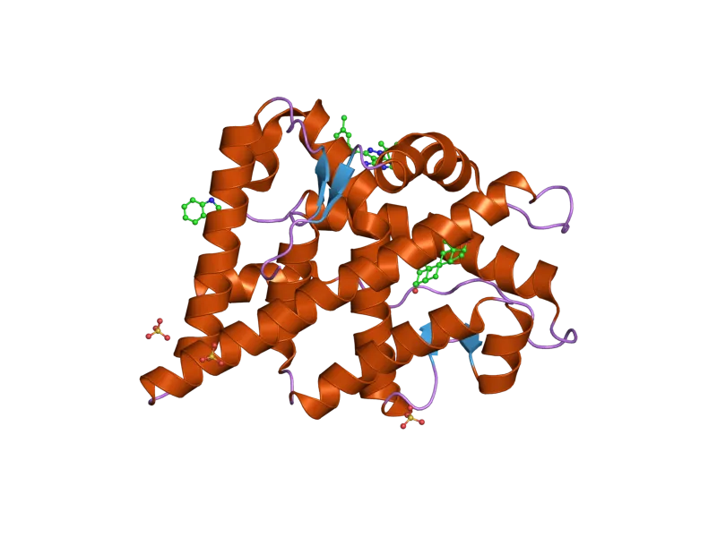
X-ray crystallography has been indispensable in decoding these interactions at atomic resolution. By crystallizing steroid-receptor complexes and diffracting X-rays through them, researchers generate electron density maps that pinpoint the position of every atom within the binding interface. These maps have revealed that steroid binding pockets are lined with hydrophobic residues that cradle the nonpolar steroid scaffold, while strategically placed polar amino acids form hydrogen bonds with the steroid's hydroxyl and ketone groups.
The androgen receptor crystal structure, first resolved in the early 2000s, demonstrated how testosterone and dihydrotestosterone occupy a predominantly hydrophobic cavity, with the C-3 ketone and C-17 hydroxyl groups forming critical hydrogen bonds with glutamine and asparagine residues, respectively. Synthetic androgens modified at the C-17 position—such as the 17-alpha alkylated compounds used historically in clinical settings—retain binding capacity while altering metabolic stability, explaining their prolonged activity compared to natural testosterone.
Crystallographic data from estrogen receptor complexes has been equally revealing. The estrogen receptor's binding pocket is notably flexible, accommodating structurally diverse ligands ranging from estradiol to selective estrogen receptor modulators. Subtle differences in how these molecules contact helix 12 determine whether the receptor acts as a transcriptional activator or repressor in a given tissue—a phenomenon known as tissue-selective agonism that has driven the development of targeted therapies for breast cancer and osteoporosis.
Carbon Numbering and the Pharmacological Consequences of Small Changes
The steroid nucleus consists of four fused rings—three cyclohexane rings and one cyclopentane ring—conventionally labeled A through D. Carbon atoms within this scaffold are numbered systematically, and the pharmacological literature routinely refers to modifications at specific positions to describe structural variants. This numbering system is not merely taxonomic; it serves as a shorthand for predicting biological behavior.
Modifications at C-6, for example, frequently enhance glucocorticoid potency. The addition of a fluorine atom at C-9 dramatically increases anti-inflammatory activity, a discovery that led to the development of fludrocortisone and later to the highly potent fluorinated corticosteroids used in inhaled asthma therapies today. Each fluorine substitution alters the electron distribution across the steroid scaffold, affecting both receptor binding affinity and resistance to enzymatic degradation.
At C-11, the presence or absence of an oxygen function determines whether a compound can activate the glucocorticoid receptor at all. Cortisone, which carries a ketone at C-11, is pharmacologically inactive until hepatic 11-beta-hydroxysteroid dehydrogenase converts it to cortisol, which bears a hydroxyl group at that position. This metabolic activation step is not incidental—it represents a biological gating mechanism that controls where and when glucocorticoid signaling occurs within the body.
Tissue Selectivity and the Problem of Off-Target Effects
One of the central challenges in steroid pharmacology is achieving tissue selectivity—delivering the desired signal to a target organ while minimizing activity elsewhere. Because the same nuclear receptor can be expressed in multiple tissues, a compound that activates the androgen receptor in muscle will also activate it in the prostate, liver, and sebaceous glands unless structural modifications are introduced to exploit differences in co-regulator expression or local enzyme activity.
Computational approaches, including molecular docking simulations and molecular dynamics modeling, have become essential tools for predicting selectivity before a compound is synthesized. These methods use the crystallographic coordinates of receptor binding domains to simulate how candidate molecules will interact with the binding pocket, estimating binding free energies and identifying which structural features favor engagement with one receptor subtype over another. While no computational model fully replicates the complexity of in vivo pharmacology, these tools have substantially accelerated the identification of promising candidates in drug discovery programs.
Why Structure Will Always Define Function
The relationship between steroid structure and biological function is not merely an academic curiosity—it is the organizing principle of an entire branch of medicine. Every synthetic corticosteroid prescribed for an autoimmune flare, every selective estrogen receptor modulator used in breast cancer management, and every androgen analog explored for muscle-wasting conditions owes its clinical profile to deliberate manipulation of molecular architecture. As structural biology and computational chemistry continue to advance, the ability to design steroid compounds with precisely targeted receptor interactions will only improve. The molecular handshake between a steroid and its receptor remains one of the most consequential interactions in human physiology, and decoding its rules continues to be among the most productive endeavors in pharmacological science.