Cortisol's Double Life: When a Life-Saving Hormone Turns Against the Body It Was Built to Protect
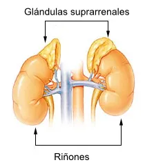
Photo: Basquetteur, arcadian, ortisa, CC0, via Wikimedia Commons
Cortisol occupies a peculiar position in human physiology. Synthesized in the adrenal cortex from cholesterol and released in response to signals from the hypothalamic-pituitary-adrenal (HPA) axis, it is simultaneously one of the most essential hormones in the body and, under the wrong conditions, one of the most damaging. Understanding why requires looking closely at the molecular architecture of its action—and recognizing that the body's stress response was never engineered for the world most Americans now inhabit.
The Architecture of an Acute Response
At baseline, cortisol circulates in a carefully regulated diurnal rhythm, peaking in the early morning hours and declining through the afternoon and evening. This rhythm is not incidental—it synchronizes metabolic activity, immune readiness, and cardiovascular tone with the demands of waking life. At these physiological concentrations, cortisol performs a suite of essential functions: it stimulates gluconeogenesis in the liver, modulates immune cell trafficking, maintains vascular responsiveness to catecholamines, and supports cognitive alertness.
When an acute stressor is encountered—a near-miss car accident, a sudden physical threat, a high-stakes presentation—cortisol levels surge within minutes. The hormone binds to glucocorticoid receptors (GRs), which are expressed in virtually every tissue in the body. Upon binding, the cortisol-GR complex translocates to the nucleus, where it functions as a transcription factor, upregulating genes involved in energy mobilization and downregulating those governing inflammation and immune activation. The result is a rapid, coordinated physiological shift toward survival readiness.
This is cortisol operating as intended: a precisely timed biochemical signal with a defined endpoint. The stressor resolves, negative feedback mechanisms engage—cortisol itself suppresses further CRH and ACTH secretion from the hypothalamus and pituitary—and concentrations return to baseline. The system resets.
Where the Design Breaks Down
The problem emerges when stressors do not resolve. Chronic occupational pressure, financial insecurity, social conflict, sleep deprivation, and the ambient anxiety generated by modern information environments all activate the HPA axis with a frequency and duration that the system was never calibrated to handle. The result is sustained cortisol elevation—not the dramatic spikes of acute stress, but a persistent upward shift in baseline that erodes the very tissues and systems the hormone was meant to protect.
At the molecular level, chronic glucocorticoid exposure produces a phenomenon known as glucocorticoid receptor downregulation. Prolonged ligand binding drives receptor internalization and reduces the transcriptional sensitivity of target tissues. This creates a paradox: the body becomes simultaneously flooded with cortisol and increasingly resistant to its regulatory signals. The anti-inflammatory and metabolic feedback loops that cortisol normally orchestrates begin to fail, even as circulating concentrations remain elevated.
Metabolic Consequences: Rewiring Energy Allocation
One of the most clinically significant consequences of chronic cortisol elevation is the profound disruption of metabolic homeostasis. Cortisol promotes gluconeogenesis and glycogenolysis to ensure glucose availability during stress. Under chronic activation, this translates into persistently elevated blood glucose, driving compensatory hyperinsulinemia. Over time, peripheral tissues—particularly skeletal muscle—develop insulin resistance, a mechanism partly mediated by cortisol's interference with insulin receptor substrate (IRS-1) signaling cascades.
Simultaneously, cortisol exerts a lipolytic effect in peripheral fat depots while paradoxically promoting adipogenesis in visceral adipose tissue. The molecular basis for this regional specificity lies in the differential expression of 11β-hydroxysteroid dehydrogenase type 1 (11β-HSD1), an enzyme that locally regenerates active cortisol from inactive cortisone. Visceral fat expresses high levels of 11β-HSD1, amplifying local glucocorticoid signaling and driving the characteristic central adiposity associated with chronic stress and conditions such as Cushing's syndrome—a state of pathological cortisol excess that serves as an informative extreme on the same biochemical continuum.
Protein catabolism represents another dimension of this metabolic dysregulation. Cortisol suppresses protein synthesis in skeletal muscle by inhibiting the mTOR pathway and upregulating the ubiquitin-proteasome degradation system. The resulting muscle wasting—sarcopenia accelerated by chronic stress—has direct implications for metabolic rate, physical function, and long-term health outcomes.
Immune Suppression vs. Immune Dysregulation
Cortisol's relationship with the immune system is frequently mischaracterized as simply immunosuppressive. In acute contexts, its anti-inflammatory actions are protective and necessary—suppressing cytokine storms and preventing the immune response from causing collateral tissue damage. Under chronic elevation, however, the picture grows considerably more complicated.
Prolonged cortisol exposure suppresses adaptive immunity by reducing lymphocyte proliferation, impairing natural killer cell activity, and shifting cytokine profiles away from the Th1 responses required for antiviral and antitumor defense. At the same time, chronic stress is associated with a paradoxical upregulation of certain pro-inflammatory pathways, partly because glucocorticoid receptor resistance prevents the normal anti-inflammatory transcriptional program from executing properly. The net result is a state of low-grade systemic inflammation combined with impaired immune surveillance—a combination that underlies elevated risk for infectious illness, autoimmune flares, and potentially cancer progression.
Neurological and Structural Damage
Perhaps the most striking evidence of cortisol's pathogenic potential under chronic conditions comes from neuroscience. The hippocampus, a brain region critical for memory consolidation and HPA axis negative feedback, expresses exceptionally high levels of glucocorticoid receptors. Sustained cortisol elevation is neurotoxic to hippocampal neurons, suppressing neurogenesis, reducing dendritic branching, and ultimately contributing to measurable volumetric loss—changes documented in individuals with major depressive disorder, post-traumatic stress disorder, and chronic anxiety conditions.
Beyond the brain, chronic cortisol exposure impairs bone mineral density by suppressing osteoblast activity and accelerating osteoclast-mediated resorption, raising long-term fracture risk. Skin integrity, collagen synthesis, and wound healing are similarly compromised through cortisol's inhibition of fibroblast proliferation and its antagonism of growth factor signaling.
Recognizing the Threshold
The central insight from this body of biochemical evidence is that cortisol's pathological effects are not a failure of the molecule itself—they are a consequence of duration and context. The same transcriptional programs that confer survival advantage in acute crisis become destructive when locked in the activated state. Clinically, this distinction matters enormously: it argues against the simplistic framing of cortisol as uniformly harmful and instead calls for precision in understanding when intervention—whether pharmacological, behavioral, or lifestyle-based—is genuinely warranted.
For clinicians and researchers, measuring not just circulating cortisol but also markers of glucocorticoid receptor sensitivity, diurnal rhythm integrity, and downstream metabolic indicators provides a more complete picture of HPA axis health than any single biomarker can offer. The science of cortisol is ultimately the science of timing—and the body, it turns out, is exquisitely sensitive to when its own chemistry runs too long.