The Androgen-to-Estrogen Conversion: Aromatase and the Hidden Feedback Architecture of Steroid Action
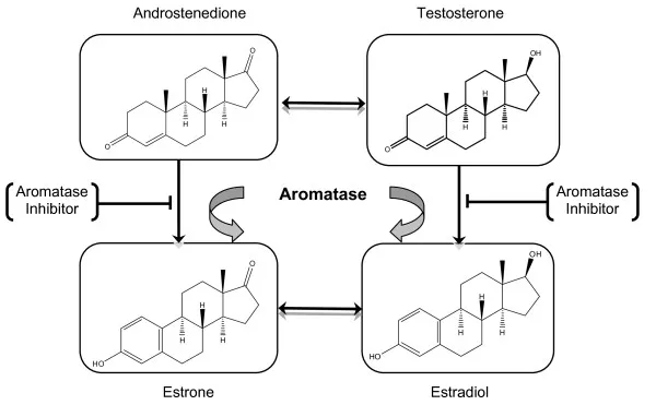
Photo: aromatase enzyme estrogen androgen hormone conversion molecular biology illustration, via cdn.ncbi.nlm.nih.gov
Steroid biochemistry rarely unfolds along a single pathway. For every androgen introduced into the body—whether endogenous or pharmaceutical—a portion of that compound is quietly redirected by an enzyme that most pharmacology discussions relegate to a footnote. Aromatase, encoded by the CYP19A1 gene, catalyzes the irreversible conversion of androgens into estrogens, and its activity reverberates through endocrine systems in ways that challenge any simple account of how steroids work.
The conventional framing of androgens and estrogens as separate, sex-specific hormones obscures a more intricate biochemical reality. Estradiol, the most biologically potent estrogen, is synthesized in significant quantities from testosterone and androstenedione in tissues distributed throughout the human body. In men and postmenopausal women alike, aromatization in peripheral tissues—rather than gonadal secretion—represents the dominant source of circulating estrogen. Understanding this conversion is not optional for steroid science; it is foundational.
The Aromatase Reaction: Chemistry at the Active Site
Aromatase belongs to the cytochrome P450 superfamily (CYP19A1) and performs one of the more structurally demanding transformations in steroid biochemistry: the conversion of the A-ring of androgen precursors from a cyclohexanone configuration to a fully aromatic phenolic ring. This aromatization process requires three sequential hydroxylation steps and the consumption of three molecules of molecular oxygen and NADPH per conversion cycle.
The substrate specificity of aromatase is notable. It accepts testosterone, androstenedione, and 16-alpha-hydroxyandrostenedione as primary substrates, converting them to estradiol, estrone, and estriol, respectively. The structural outcome—a phenolic A-ring—fundamentally alters the hormone's receptor binding profile, shifting affinity away from the androgen receptor and toward estrogen receptor alpha and beta isoforms. This is not a minor modification; it represents a complete reclassification of the molecule's biological identity.
The enzyme's active site geometry also makes it a tractable pharmacological target. Aromatase inhibitors—competitive and mechanism-based—have been developed with considerable precision, and their clinical deployment in oncology and endocrinology has generated an extensive body of research illuminating how consequential this single enzymatic step truly is.
Tissue-Specific Expression: Aromatase Is Not Confined to the Gonads
One of the most consequential features of aromatase biology is its distribution across tissues that were historically not considered primary sites of steroid synthesis. While gonadal aromatization is well established, the enzyme is also expressed in adipose tissue, bone, the brain, skin, and vascular endothelium—each of these sites capable of locally converting circulating androgens into estrogens that act in paracrine or autocrine fashion.
Adipose tissue deserves particular emphasis in a US clinical context, given the epidemiological significance of obesity in American patient populations. Adipocytes express aromatase constitutively, and enzyme activity scales with adipose mass. Individuals with greater adiposity therefore exhibit elevated rates of peripheral aromatization, producing higher circulating estrogen levels from any given androgen substrate—whether that substrate originates endogenously or from therapeutic administration. This relationship has direct implications for the clinical management of testosterone replacement therapy, where adiposity is a meaningful predictor of estrogenic side effects.
In bone tissue, local aromatization plays an essential role in skeletal maintenance for both sexes. Estrogen derived from androgen precursors regulates osteoclast activity and bone mineral density through estrogen receptor signaling in osteoblasts. The clinical significance of this pathway became dramatically apparent in case studies of men with aromatase deficiency or estrogen receptor mutations, who develop severe osteoporosis despite normal or elevated androgen levels—demonstrating that androgens alone cannot maintain bone mass without local estrogenic conversion.
Brain aromatization represents perhaps the most underappreciated dimension of this story. Neurons in the hypothalamus and limbic system express aromatase, and locally produced estradiol modulates synaptic plasticity, neuroprotective responses, and the regulation of the hypothalamic-pituitary-gonadal (HPG) axis itself.
The HPG Axis Feedback Loop: Estrogen as the Primary Signal
A common misconception in lay discussions of androgen biology is that testosterone itself is the primary negative feedback signal at the level of the hypothalamus and pituitary. The research literature tells a more nuanced story. Estradiol—produced via aromatization of testosterone—exerts substantially more potent negative feedback on gonadotropin-releasing hormone (GnRH) pulsatility and on luteinizing hormone (LH) and follicle-stimulating hormone (FSH) secretion than testosterone does directly.
Studies using selective estrogen receptor modulators and aromatase inhibitors in male subjects have consistently demonstrated that blocking estrogenic signaling—even while maintaining androgen levels—produces significant increases in LH and FSH, confirming that the HPG axis is reading estrogen as its principal feedback signal. This has profound implications for understanding how exogenous androgen administration suppresses the HPG axis: the suppression operates largely through aromatization-derived estradiol, not through androgen receptor activation at the pituitary alone.
This feedback architecture also explains why aromatase inhibitors can serve as tools to stimulate endogenous testosterone production in certain clinical protocols. By reducing the estrogenic signal at the hypothalamus and pituitary, these agents partially disinhibit GnRH and gonadotropin secretion, resulting in elevated LH drive to the testes and increased testosterone output—a mechanism that has found application in the management of hypogonadism and in fertility medicine.
Clinical Consequences of Uncontrolled Aromatization
The physiological consequences of excessive aromatase activity extend well beyond reproductive endocrinology. Elevated estradiol in men has been associated with gynecomastia—proliferation of glandular breast tissue driven by estrogen receptor activation—as well as with alterations in fluid balance, lipid metabolism, and cardiovascular risk markers. In clinical settings involving therapeutic androgen administration, aromatization management has become a recognized component of treatment protocol design.
Conversely, aromatase deficiency—whether congenital or pharmacologically induced—carries its own pathological burden. Beyond the skeletal consequences described above, estrogen insufficiency in both sexes is associated with metabolic dysregulation, including impaired insulin sensitivity and adverse lipid profiles. The assumption that minimizing estrogen is unconditionally beneficial has been substantially revised by the clinical literature, which increasingly supports the view that estrogen, even in male physiology, operates within an optimal range rather than functioning as a purely undesirable byproduct.
Breast cancer biology has provided some of the most rigorous data on aromatase function, as the development of third-generation aromatase inhibitors for estrogen receptor-positive malignancies required precise characterization of the enzyme's activity across tissue compartments. The oncology literature has, in this sense, inadvertently furnished steroid science with some of its most detailed mechanistic insights.
Aromatization as a Design Principle in Steroid Pharmacology
Synthetic steroid development has long grappled with aromatization as both a problem to be solved and a property to be preserved depending on therapeutic context. Structural modifications that reduce or eliminate aromatase substrate recognition—such as fluorine substitution at C2 or modifications to the A-ring—have been incorporated into compounds where estrogenic activity is undesirable. Understanding which structural features confer or prevent aromatase recognition remains an active area of steroid chemistry research.
What the aromatization pathway ultimately illustrates is that steroids do not act in pharmacological isolation. Every androgen introduced into the body initiates a branching network of enzymatic responses, receptor interactions, and feedback signals—many of which operate through estrogen, not androgen, signaling. Treating aromatization as a secondary consideration rather than a primary architectural feature of steroid biochemistry leads to an incomplete and sometimes misleading picture of how these compounds behave in living systems.